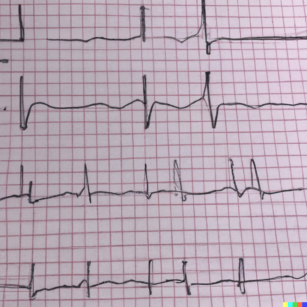
An electrocardiogram (ECG) is a test that measures the electrical activity of the heart. It records the heart’s rhythm and activity on a moving strip of paper or a digital display. It is used to diagnose a variety of cardiac conditions, including heart attacks, arrhythmias, and heart failure.
The ECG records the electrical activity of the heart as waves. The waves are labeled P, Q, R, S, and T. The P wave represents the electrical activity that causes the atria (the upper chambers of the heart) to contract. The Q, R, and S waves represent the electrical activity that causes the ventricles (the lower chambers of the heart) to contract. The T wave represents the electrical activity that causes the ventricles to relax.
There are several parameters that are commonly measured on an ECG, including:
- Heart rate: The number of times the heart beats per minute.
- P-R interval: The time between the start of the P wave and the start of the QRS complex.
- QRS duration: The time between the start of the Q wave and the end of the S wave.
- Q-T interval: The time between the start of the Q wave and the end of the T wave.
The ECG can also be used to diagnose specific conditions such as:
- Atrial fibrillation (AF) : A condition in which the atria contract rapidly and irregularly.
- Myocardial infarction (MI) : heart attack, caused by a blockage of blood flow to the heart muscle.
- Bundle branch block (BBB) : caused by a problem with the electrical pathways of the heart.
- Heart failure: A condition in which the heart is unable to pump enough blood to meet the body’s needs.
An ECG is a non-invasive and painless procedure that can be performed quickly in a doctor’s office or hospital setting. The results are usually available within minutes and interpreted by a healthcare professional.
Leave a Reply