The cardiac cycle is the series of events that occur during a single heartbeat, from the beginning of one contraction of the heart to the beginning of the next. It is composed of several phases, each with its own characteristics and functions.
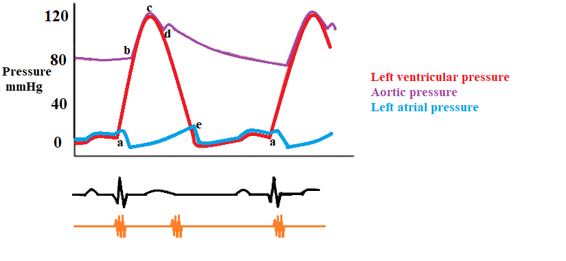
The first phase is called the isovolumetric ventricular contraction (a-b), and it marks the beginning of systole. This phase starts with the appearance of the QRS complex on the electrocardiogram (EKG) and the closure of the atrioventricular (AV) valves at point (a). During this phase, the ventricle generates positive pressure without any change in its volume, which is referred to as isovolumetric. This helps to overcome the resistance of the semilunar valves and eventually causes them to open at point (b). This phase lasts approximately 6% of the cardiac cycle.
The next phase is the rapid ejection (b-c), during which the semilunar valves open at point (b) and blood is rapidly ejected from the ventricles due to increased contractility. As a result, arterial pressure increases until reaching its maximum at point (c). This phase lasts for about 13% of the cardiac cycle.
The reduced ejection (c-d) phase marks the beginning of ventricular repolarization, which is depicted by the onset of the T wave on the EKG. Repolarization leads to a rapid decline in ventricular pressures, resulting in a reduced rate of ejection. Despite this decline, some forward flow of blood continues due to the remnants of kinetic energy from the previous phase. This phase lasts approximately 15% of the cardiac cycle.
The isovolumetric relaxation (d-e) phase begins when the ventricular pressures drop below the diastolic aortic and pulmonary pressures, causing the aortic and pulmonary valves to close and producing the second heart sound (point d). This marks the beginning of diastole, during which the ventricles generate negative pressure without changing their volume, which helps to lower the ventricular pressure below the atrial pressure. This phase lasts for about 8% of the cardiac cycle.
The ventricular filling (e-a) phase starts when the AV valves open at point (e), allowing the ventricles to fill with blood. The initial rapid filling is mainly due to ventricular suction, which is caused by the return of each ventricular muscle fiber to its slack length and the ventricular untwisting. The ventricular pressure gradually increases until it equals the atrial pressure, causing the AV valves to close at point (a). This phase lasts for approximately 44% of the cardiac cycle.
Finally, near the end of ventricular diastole, atrial contraction contributes about 10% of the ventricular filling volume, which is represented by the P wave on the EKG of the following cycle. This phase lasts for about 14% of the cardiac cycle.
In conclusion, the cardiac cycle is a complex series of events that occur during each heartbeat, allowing the heart to pump blood effectively and efficiently throughout the body. Understanding the different phases of the cardiac cycle is crucial for understanding the functioning of the heart and diagnosing any issues that may arise.